An MRI giant to explore the depths of the brain

sudok1

Published the 08.07.2017 at 13: 15


A A

Keywords :
IRMneurologieinnovation
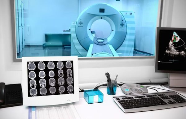
Five meters of diameter and five meters long. Here are the measurements of the largest tool resonance Imaging (mri) medical (MRI), the center for research in neuroimaging brain Neurospin, based in Saclay (Essonne) has been available to celebrate its ten years of existence.
Presented on July 6 at the press, this giant magnet is unique in the world requires the intervention of a team of over 170 researchers for its implementation in the road.
To 11.7 tesla
The device has been designed in partnership with the project of a franco-German Isolde, a collaboration between the CEA, Guerbet (manufacturer of contrast agents for imaging), university of Freiburg (Germany) and the manufacturer Siemens.
It took 10 years to shape this giant magnet of 11.7 teslas (unit of measure of magnetic induction). A power almost four times greater than that of MRI scanners standard 1.5 to 3 tesla, which allow you to observe millions of neurons.
“At the other end of the spectrum, we also have to NeuroSpin, for the research, an MRI scanner 17 teslas, which lets you see the neurons individually in the animal. We lacked an intermediate scale, ” says Denis Le Bihan, founder and director of NeuroSpin.
A better understanding of es neurological pathologies
According to the scientists, this new field of exploration of the brain would allow to study the cerebral cortex in depth, the thin layer of tissue that houses the cognitive functions of the human brain. In 2016, the results of a study by MRI at 3 T of a american consortium of the Human Connectome Project have shown that areas of the cortex were divided into 200 areas, each corresponding to a cognitive function.
Denis Le Bihan intends to go even further in discovering other regions of the brain (also known as “areas of Brodmann), in order to better understand the neurological pathologies or psychiatric disorders such as Alzheimer’s, epilepsy or schizophrenia.
“With this instrument, the vision of the brain could radically change. Thanks to the high-resolution images, we hope to see the first amyloid plaques characteristic of Alzheimer’s disease. This would allow for a diagnosis very early, ” hopes Denis Le Bihan.
First trial on patients in 2019
But before you achieve such results, the MRI 11.7 T has a long way to go. Once started, its magnetic field will be increased in a phased manner, in order to verify that everything works correctly.
When this step is approved, the institute NeuroSpin must obtain the authorization of the national security Agency of medicines and health products (MSNA) to test the machine on patients. This procedure may take several years, the team of the CEA hopes to get the green light, ” by 2019 “.
YOU MAY ALSO LIKE
 International Software Engineering Olympiad PROD Launches 2025–2026 Season
The world’s first high-school competition focused on application development and real business processes of major tech companies ...
READ MORE A Man’s Death Was Reported As A Possible Bear Mauling. Police Say It Was Murder
Montana authorities say a 35-year-old man camping in a remote forest was found brutally killed in his tent.
A 35-year-old man ...
READ MORE Cameron Diaz Explains Why Nothing ‘Could Change My Mind’ About Retiring In 2018
The "Charlie's Angels" star last appeared on-screen in 2014 and, despite an impending comeback, explained Monday why quitting "felt ...
READ MORE JoJo Siwa Opens Up About The ‘Brutal’ Backlash She Received For Controversial Magazine Cover
Siwa compared the look to Harry Styles’ viral 2020 cover in Vogue.
JoJo Siwa is getting candid about the “brutal” backlash she has ...
READ MORE UN expert on violence against women and girls takes shot at IOC over women’s boxing
The International Olympic Committee under president Thomas Bach has sought to work closely with the United Nations. Particularly when ...
READ MORE The February Artist of the Month selected in the BE OPEN Regional Art competition for emerging artists
BE OPEN Art, an online art gallery set up by BE OPEN think tank, a humanitarian initiative founded by international entrepreneur and ...
READ MORE